Histology
Real, openly licensed radiology, histology and pathology images with teaching points. Zoom, pan and adjust each image in your browser.
! Sample content, not yet medically reviewed. This page is not indexed.
For education only. Not medical advice and not for diagnosis or treatment decisions.
Educational use only. These images are not for diagnosis, treatment or any clinical decision-making.
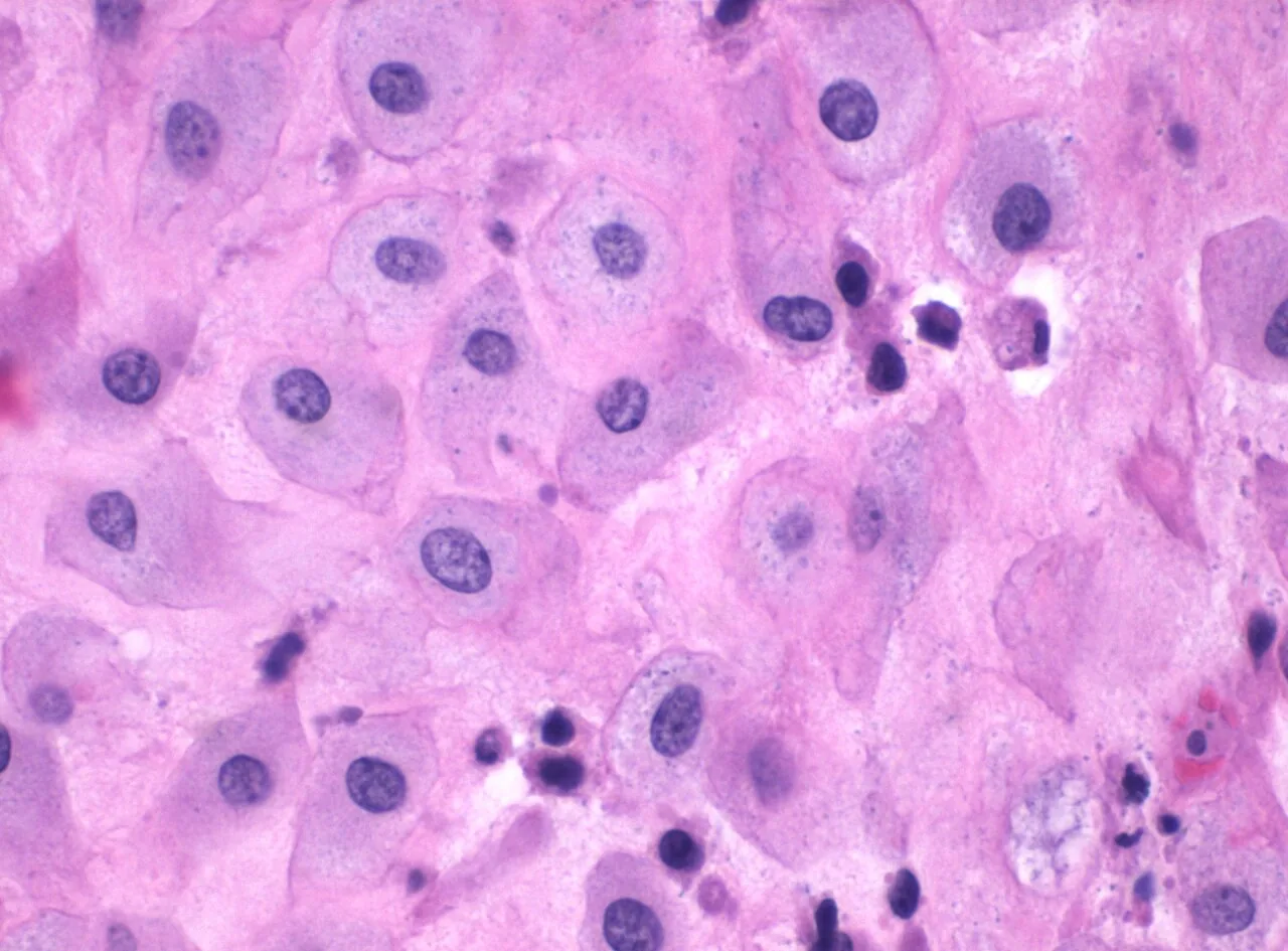
Decidua (H&E, 400x)
Normal example
Haematoxylin and eosin stain at 400x, from the products of a miscarriage. Decidua is the pregnancy-modified lining of the uterus.
Educational use only. These images are not for diagnosis, treatment or any clinical decision-making.
 Zoom 100%
Drag the image to move it. With the image focused, use the arrow keys to pan and + / - to zoom.
Credit: Image by Mikael Häggström, M.D. · Licence: CC0 1.0 · Source: Wikimedia Commons What to look at
- Haematoxylin stains nuclei blue-purple and eosin stains cytoplasm and collagen pink.
- Decidual cells are large and polygonal with abundant pink cytoplasm and well-defined cell borders.
- Nuclei are round, centrally placed and similar in size from cell to cell.
- The cells sit in sheets, closely packed, which gives the tissue its uniform pink look at this magnification.
- A few small dark nuclei between the large cells are other cells in the stroma, such as lymphocytes.
! Teaching points are AI-drafted and have not been reviewed by a clinician.
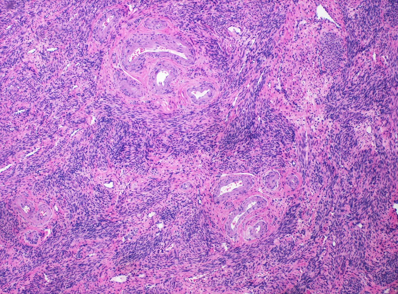
Postmenopausal myometrium (H&E, low power)
Normal example
Haematoxylin and eosin stain at low magnification of the muscular wall of the uterus after the menopause; the source notes that some blood vessels are visible.
Educational use only. These images are not for diagnosis, treatment or any clinical decision-making.
 Zoom 100%
Drag the image to move it. With the image focused, use the arrow keys to pan and + / - to zoom.
Credit: Image by Mikael Häggström, M.D. · Licence: CC0 1.0 · Source: Wikimedia Commons What to look at
- Low power is for pattern: look first at how the tissue is arranged before looking at single cells.
- Smooth muscle is arranged in interlacing bundles, so some bundles are cut lengthwise and others across.
- Smooth muscle cells have eosinophilic (pink) cytoplasm and elongated, cigar-shaped nuclei.
- Blood vessels with thicker walls and a visible lumen run between the bundles.
- Blue-purple areas are denser in nuclei, usually more cellular regions or small collections of other cells.
! Teaching points are AI-drafted and have not been reviewed by a clinician.
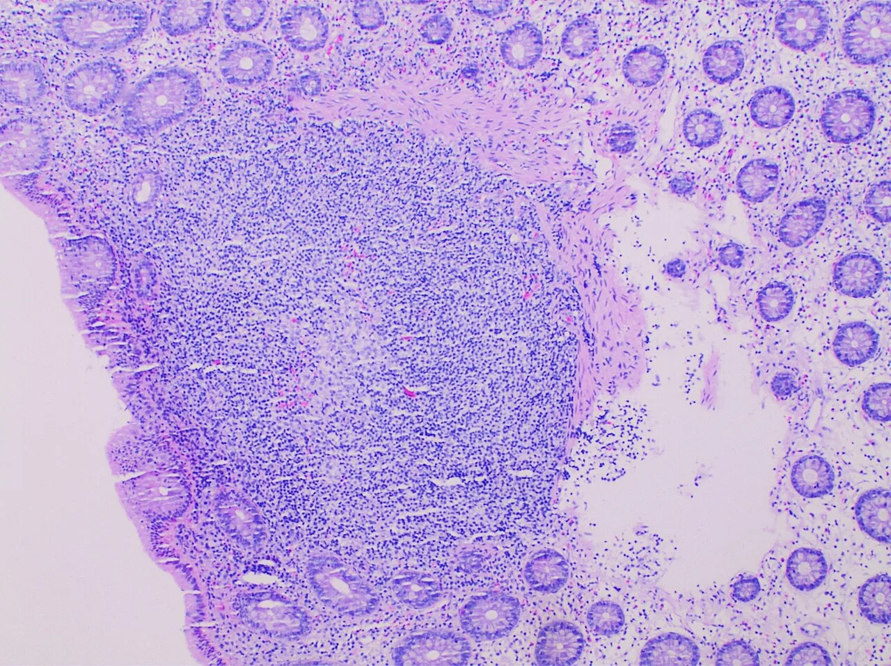
Colon mucosa with a lymphoid aggregate (H&E)
Normal example
Haematoxylin and eosin stain of a subepithelial lymphoid aggregate in the colon. Lymphoid tissue in the gut wall is a normal part of the immune system of the mucosa.
Educational use only. These images are not for diagnosis, treatment or any clinical decision-making.
 Zoom 100%
Drag the image to move it. With the image focused, use the arrow keys to pan and + / - to zoom.
Credit: Image by Mikael Häggström, M.D. · Licence: CC0 1.0 · Source: Wikimedia Commons What to look at
- The colon mucosa has straight, closely packed crypts, seen here as round or elongated pale profiles lined by epithelium.
- Goblet cells give the crypt lining a pale, bubbly look because their mucus is washed out in processing.
- The dense blue-purple area is made of small round cells with dark nuclei and little cytoplasm, typical of lymphocytes.
- The aggregate sits just beneath the surface epithelium, in the lamina propria, and pushes the crypts apart.
- Lymphoid aggregates are a normal part of the gut-associated lymphoid tissue, so this is normal anatomy.
! Teaching points are AI-drafted and have not been reviewed by a clinician.
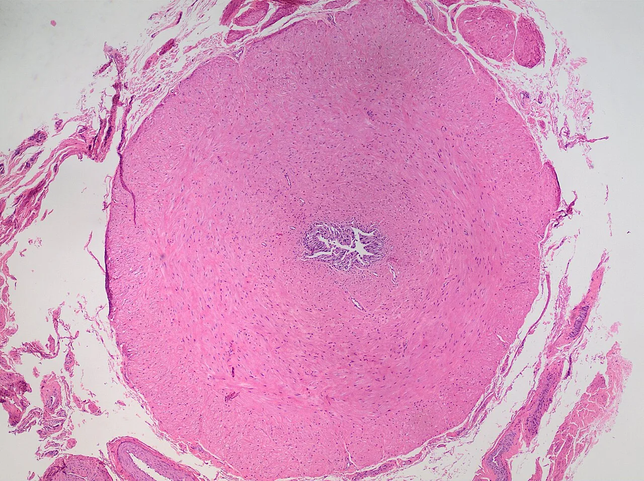
Vas deferens in cross-section (H&E)
Normal example
Cross-section of an unremarkable vas deferens, haematoxylin and eosin stain. The duct carries sperm from the epididymis toward the urethra.
Educational use only. These images are not for diagnosis, treatment or any clinical decision-making.
 Zoom 100%
Drag the image to move it. With the image focused, use the arrow keys to pan and + / - to zoom.
Credit: Image by Mikael Häggström, M.D. · Licence: CC0 1.0 · Source: Wikimedia Commons What to look at
- The wall is very thick compared with the lumen: most of the circle is smooth muscle, which is why the duct can be felt as a firm cord.
- The lumen is narrow and star-shaped because the mucosa is folded into longitudinal ridges.
- The lining is a tall pseudostratified epithelium on a thin connective tissue layer.
- The muscle coat is arranged in layers of different fibre direction: inner and outer longitudinal and a middle circular layer.
- Surrounding connective tissue and small vessels lie outside the muscle, with nerves in the outer layer.
! Teaching points are AI-drafted and have not been reviewed by a clinician.


Related
Clinical cases
Step-by-step cases with decisions and feedback. AI-drafted, not yet medically reviewed.
Interactive 3D anatomy atlas
Rotate and isolate body structures by system. Names are not yet medically reviewed.