MRI
Real, openly licensed radiology, histology and pathology images with teaching points. Zoom, pan and adjust each image in your browser.
! Sample content, not yet medically reviewed. This page is not indexed.
For education only. Not medical advice and not for diagnosis or treatment decisions.
Educational use only. These images are not for diagnosis, treatment or any clinical decision-making.
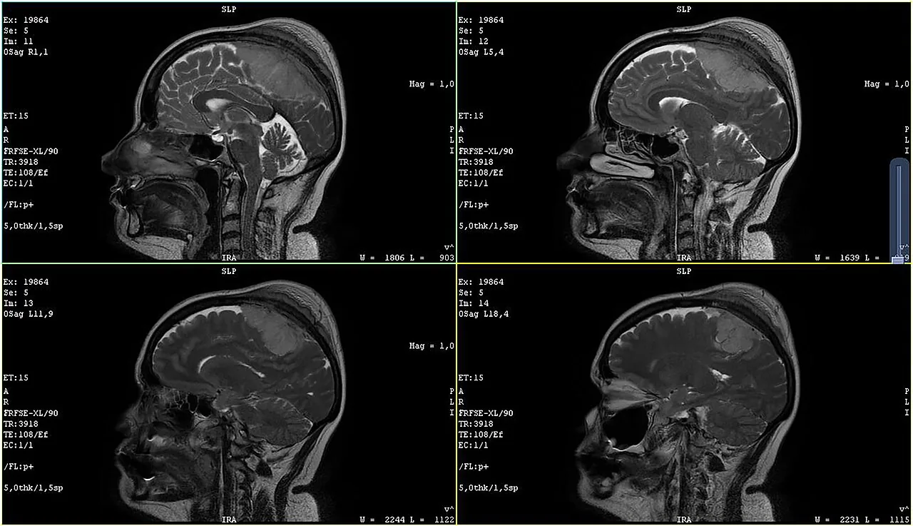
Sagittal brain MRI series (source: meningioma case)
Example with a finding described by the source
Four sagittal slices of a brain MRI. The source describes a meningioma in a 52-year-old woman; this page uses the images to teach MRI appearances, not to make or discuss a diagnosis.
Educational use only. These images are not for diagnosis, treatment or any clinical decision-making.
 Zoom 100%
Drag the image to move it. With the image focused, use the arrow keys to pan and + / - to zoom.
Credit: Image by RASPBERRY VIBE · Licence: CC0 1.0 · Source: Wikimedia Commons What to look at
- A sagittal slice cuts the head from front to back, so the midline structures are seen in profile: the corpus callosum, brainstem and cerebellum.
- On this type of image the cerebrospinal fluid is bright, grey matter is intermediate and white matter is darker.
- Cortical bone and air give almost no signal and look black, which is why the skull and sinuses look so different from CT.
- Compare slices from one side to the other: each slice is a different distance from the midline.
- The scanner text at the edges lists the series, slice number and technique. The source states there is a lesion; learners should compare the slices and ask what differs from the expected anatomy.
! Teaching points are AI-drafted and have not been reviewed by a clinician.
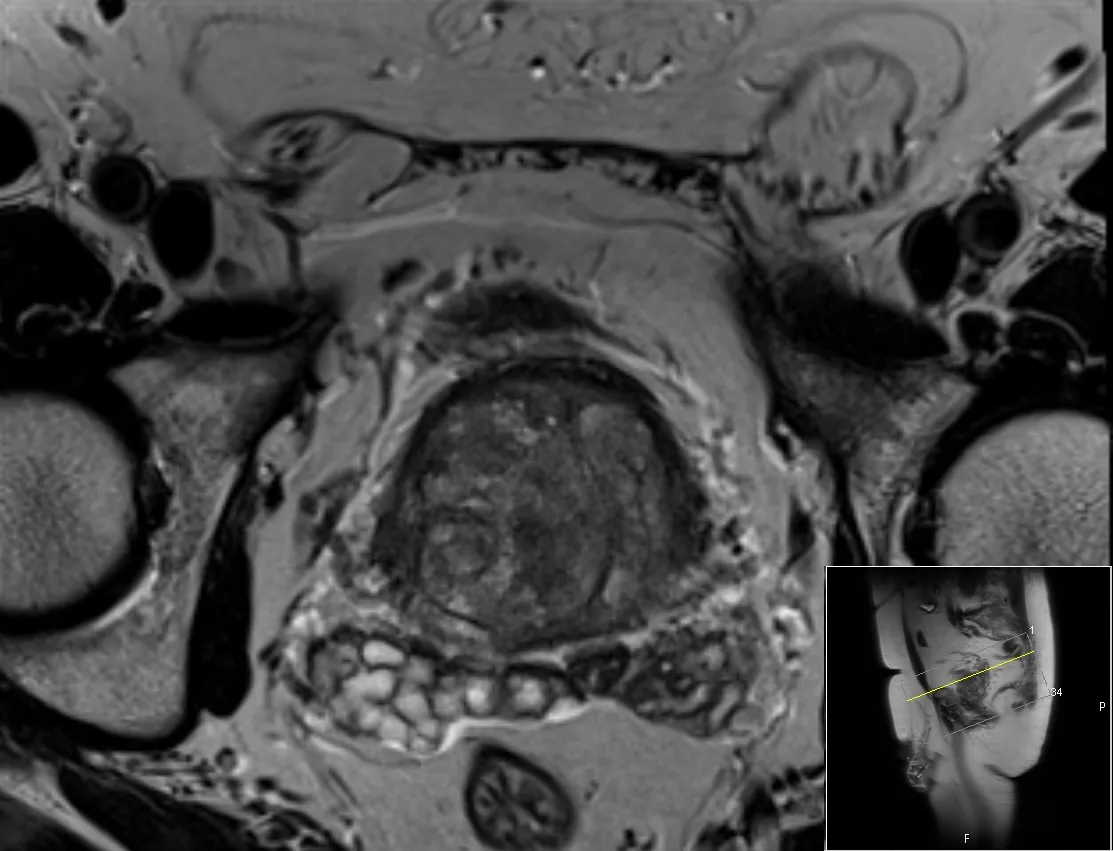
T2-weighted MRI, axial pelvis (source: inguinal hernia case)
Example with a finding described by the source
T2-weighted MRI of a 67-year-old man. The source states it shows a fat-containing left inguinal hernia (on the right side of the image). It is used here to show how an axial pelvic MRI is organised.
Educational use only. These images are not for diagnosis, treatment or any clinical decision-making.
 Zoom 100%
Drag the image to move it. With the image focused, use the arrow keys to pan and + / - to zoom.
Credit: Image by Mikael Häggström, M.D. · Licence: CC0 1.0 · Source: Wikimedia Commons What to look at
- On T2-weighted images fluid is bright and cortical bone is dark; the rounded structures at the left and right edges are the heads of the femurs.
- MRI separates soft tissues well: muscle, fat and the pelvic organs have different grey levels and clear boundaries.
- In the midline, the pelvic organs lie in front of the rectum, which is seen toward the bottom of the image.
- As on CT, the patient's right is on the image left; the inset shows the angle of the slice on a sagittal image.
- The source's finding sits at the groin on one side; asymmetry between left and right is a useful clue to look for.
! Teaching points are AI-drafted and have not been reviewed by a clinician.


Related
Clinical cases
Step-by-step cases with decisions and feedback. AI-drafted, not yet medically reviewed.
Interactive 3D anatomy atlas
Rotate and isolate body structures by system. Names are not yet medically reviewed.