CT
Real, openly licensed radiology, histology and pathology images with teaching points. Zoom, pan and adjust each image in your browser.
! Sample content, not yet medically reviewed. This page is not indexed.
For education only. Not medical advice and not for diagnosis or treatment decisions.
Educational use only. These images are not for diagnosis, treatment or any clinical decision-making.
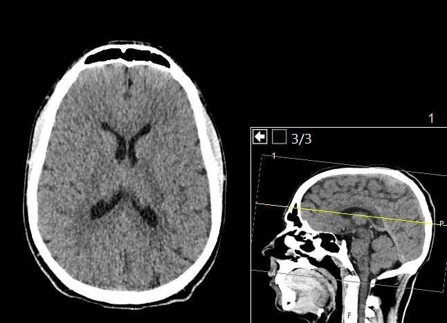
Normal head CT, axial, level of the lateral ventricles
Normal example
Non-contrast CT of the head of an 18-year-old male, 4 mm slices; the source states it shows normal anatomy with no injuries. The small inset shows the level of the slice on a sagittal image.
Educational use only. These images are not for diagnosis, treatment or any clinical decision-making.
 Zoom 100%
Drag the image to move it. With the image focused, use the arrow keys to pan and + / - to zoom.
Credit: Image by Mikael Häggström, M.D. · Licence: CC0 1.0 · Source: Wikimedia Commons What to look at
- On CT, dense structures are white (bone), air is black, and the brain is grey. Cerebrospinal fluid is darker than brain tissue.
- The grey matter of the cortex is slightly brighter than the white matter beneath it.
- The lateral ventricles are the dark, symmetrical, butterfly-shaped spaces near the middle; the midline falx divides the two hemispheres.
- Images are shown as if you were looking from the patient's feet: the patient's right is on the image left.
- The inset sagittal image shows where the slice was taken. Scrolling through slices is how a real study is read.
! Teaching points are AI-drafted and have not been reviewed by a clinician.
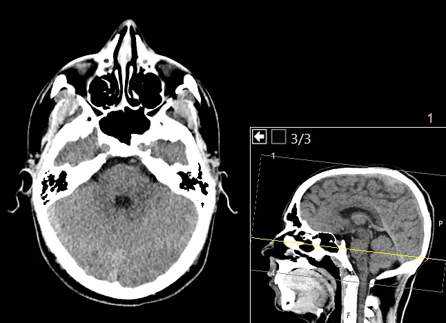
Normal head CT, axial, skull base and posterior fossa
Normal example
A lower slice from the same normal non-contrast head CT. It shows the structures near the skull base, where bone and air make the picture very different from the upper slices.
Educational use only. These images are not for diagnosis, treatment or any clinical decision-making.
 Zoom 100%
Drag the image to move it. With the image focused, use the arrow keys to pan and + / - to zoom.
Credit: Image by Mikael Häggström, M.D. · Licence: CC0 1.0 · Source: Wikimedia Commons What to look at
- Air-filled spaces are black: the nasal cavity and sinuses at the front and the mastoid air cells beside the petrous bone.
- The posterior cranial fossa at the back contains the cerebellum, and a small dark spot near its centre corresponds to the fourth ventricle region.
- Bone is white and thick near the skull base, with a complex shape, so slices here look more irregular than higher up.
- Compare left and right: symmetry is the first check for most head CT slices.
- Beam-hardening streaks near dense bone are a common technical feature at this level.
! Teaching points are AI-drafted and have not been reviewed by a clinician.
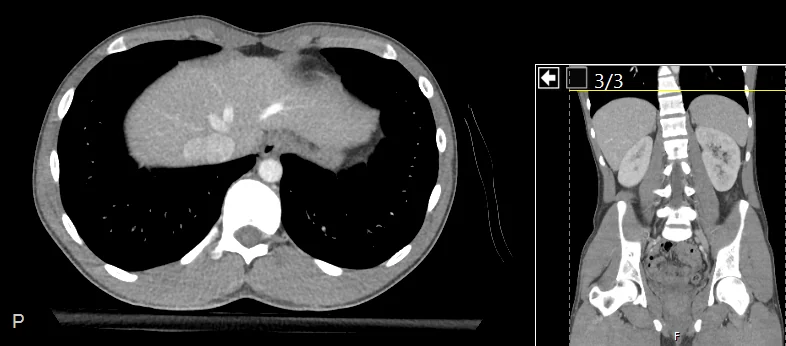
Normal contrast CT, axial, upper abdomen and lung bases
Normal example
Contrast-enhanced CT of the abdomen and pelvis of a 21-year-old male (3 mm slices); the source states the anatomy is normal. This slice is high, through the liver and the lung bases.
Educational use only. These images are not for diagnosis, treatment or any clinical decision-making.
 Zoom 100%
Drag the image to move it. With the image focused, use the arrow keys to pan and + / - to zoom.
Credit: Image by Mikael Häggström, M.D. · Licence: CC0 1.0 · Source: Wikimedia Commons What to look at
- The liver fills the upper abdomen on this slice; bright branching lines inside it are vessels filled with injected contrast.
- Both lung bases appear black (air) with a few fine grey vessel markings, lying beside and behind the liver.
- The aorta is the round bright structure just in front of the vertebral body, slightly to the patient's left.
- The vertebral body and spinal canal sit in the midline at the back, surrounded by the ribs.
- The small coronal image shows the level; in the standard convention the patient's right is on the image left.
! Teaching points are AI-drafted and have not been reviewed by a clinician.


Related
Clinical cases
Step-by-step cases with decisions and feedback. AI-drafted, not yet medically reviewed.
Interactive 3D anatomy atlas
Rotate and isolate body structures by system. Names are not yet medically reviewed.