Pathology
Real, openly licensed radiology, histology and pathology images with teaching points. Zoom, pan and adjust each image in your browser.
! Sample content, not yet medically reviewed. This page is not indexed.
For education only. Not medical advice and not for diagnosis or treatment decisions.
Educational use only. These images are not for diagnosis, treatment or any clinical decision-making.
Gross pathology: subarachnoid haemorrhage
Example with a finding described by the source
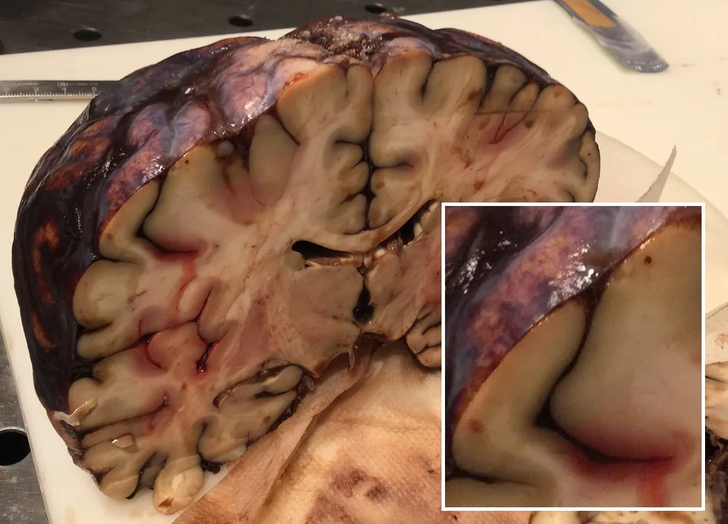
Autopsy specimen from a fatal acute subarachnoid haemorrhage, cut coronally; the arachnoid mater was left in place on the surface. The source states the blood also fills the sulci (see the magnified inset).
Educational use only. These images are not for diagnosis, treatment or any clinical decision-making.
 Zoom 100%
Drag the image to move it. With the image focused, use the arrow keys to pan and + / - to zoom.
Credit: Image by Mikael Häggström, M.D. · Licence: CC0 1.0 · Source: Wikimedia Commons What to look at
- The subarachnoid space lies between the arachnoid mater and the pia mater, so blood here coats the brain surface and follows the sulci.
- In a coronal slice the cortex is the thin outer layer, with the paler white matter inside and the pattern of gyri and sulci at the edge.
- Dark red to black material on the surface and deep in the grooves, in the magnified inset, is the blood.
- Gross pathology means looking at the whole organ without a microscope: note colour, shape, distribution and symmetry.
- This is a specimen from an autopsy case; the cause of such bleeding cannot be seen from this photograph.
! Teaching points are AI-drafted and have not been reviewed by a clinician.
Gross pathology: fresh papillary muscle infarction in the left ventricle
Example with a finding described by the source
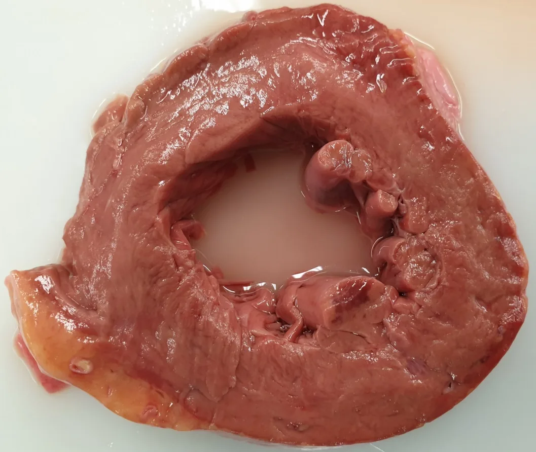
Unfixed, unstained cross-section of the heart. The source describes a fresh infarct as a 12 mm dark area in a papillary muscle of the anterior wall, and a wall about 20 mm thick (normal up to about 10 mm).
Educational use only. These images are not for diagnosis, treatment or any clinical decision-making.
 Zoom 100%
Drag the image to move it. With the image focused, use the arrow keys to pan and + / - to zoom.
Credit: Image by Mikael Häggström, M.D. · Licence: CC0 1.0 · Source: Wikimedia Commons What to look at
- A short-axis slice of the left ventricle shows a ring of thick muscle (myocardium) around the central blood-filled chamber.
- Papillary muscles are the muscular columns that project into the chamber and anchor the heart valve cords.
- Fresh infarcted muscle can look dark or mottled, but early change is often subtle on gross inspection.
- Wall thickness is a measurement: the source gives about 20 mm here versus up to about 10 mm in a normal left ventricle.
- Gross findings are matched with the history and with microscopic examination; a photograph alone does not establish a cause.
! Teaching points are AI-drafted and have not been reviewed by a clinician.


Related
Clinical cases
Step-by-step cases with decisions and feedback. AI-drafted, not yet medically reviewed.
Interactive 3D anatomy atlas
Rotate and isolate body structures by system. Names are not yet medically reviewed.